 |
|
 |
     |
Histology The microscopic study of the structure of tissue using special technique is define as Histology. The word “histology” comes from the Greek word histo which means “tissue” and logia which means "study”. Histology is considered as an essential tool of biology since it studies the microscopic structure of cell of plants and animals. The five main stages in preparing histology slides are, Fixing, Processing, Embedding, Sectioning and Staining. Histology stains depends on the tissue to be observed. The most widely used stain in histology is the Hematoxylin which stains the nuclei and Eosin which stains the cytoplasm of cells pink. 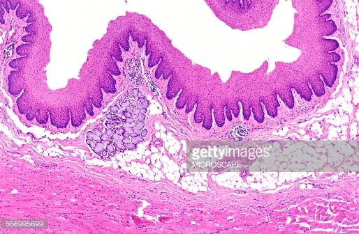 Histologists are the people who specialized in processing the tissues in histology laboratories. Histology is very important especially in; Education, in which the stained slides can be used to help the student in identifying the microstructures of cell or tissue. Diagnosis, which enables the health professional to determine the diseased cell or tissue. And autopsy, to identify the possible circumstances or cause of death by studying the biological tissue of deceased person or animal. The microscopic study of diseased biological tissue is called histopathology (a sub-discipline within pathology). Medical specialists who study and interpret diseased tissues in microscopic detail are called Histopathologists (IvyRose, 2003). 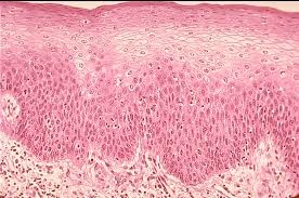 |
||
Copyright © 2016 Magic Flakes |
|||