 |
|
 |
Clinical Microscopy (Analysis of Urine and Body Fluids) One of the sections in the field of Medical technology that is usually performed in the Laboratory. It deals with the different body fluids of human being but more on urinalysis. Urinalysis is a kind of screening test that is utilized to evaluate the urine sample. It is used to diagnose or to examine variety of diseases such as urinary tract infection, kidney diseases, liver diseases and also diabetes. In the history of urinalysis, early physicians used physical examination of urine in order to diagnose different illness. They rely on the turbidity, volume and color of the urine, for example one of their basis in diagnosing is that if the color of the urine is different from the usual color they assumed that the sample is pathologic. Also the ant tasting is one of the method which usually they performed. They use ants to diagnose diabetes whether the ant is attracted to the sample or not.  Now in the 21st century the examination of urine or urinalysis have taken a huge leap in every aspect of diagnosing illnesses. More discoveries and more tools are being used to assist the health care professionals in performing rapid and accurate tests. For example, the physical examination of urine used to rely basically on the appearance of a urine sample, regardless of the way it was collected, amount of contamination it was exposed to, container used or where it was stored; in which it could be affecting the urine sample by various factors that will give a false diagnosis. But nowadays we have strict procedures and instructions on the best methods to collect the urine depending on what type of tests we are performing, assuring least amount of contamination occurring and also different types of preservatives to sustain the urine viability. Not to mention utilization of instruments such as urinometer and refractometer. Also Chemical examination, in the past times chemical examination was not a very popular test, even though it is definitely very significant in urine testing that could yield many parameters of urine that holds different interpretations for our bodily functions and their current states if normal or abnormal. Parameters such as Blood, presence of blood in urine holds a major health issue that could be caused by many factors from infections to internal bleeding or damaged kidneys. Also other parameters such as glucose and protein, their presence in urine is extremely significant since it should be reabsorbed by the body, their presence indicates a serious kidney damage that needs to be resolved. And to perform these chemical examination tests, currently there are two ways, wet lab procedures and Reagent test strip method. Wet lab procedures which is more complicated and requires different reagents, miscalculations or wrong storage could lead to false results. And the second way to perform a chemical examination is Reagent test strip method, which is the fastest and simplest way. The reagent test strip comprises of 10 chemical pads or reagents which react (change color) once immersed for a few seconds in urine and then removed. And it can be interpreted between 60 to 120 seconds. These different colors that appear on the 10 chemical pads, are compared on a colored scale provided by the manufacturer that will be used to interpret the results. 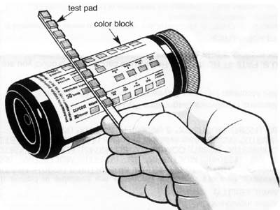 Final part of urinalysis is Microscopic examination, which is the most significant and most important exam for urine among the others for it will give a concrete result on the vital state of the body. And with the advancement of present-day technology this procedure is becoming easier and easier. It utilizes a drop of urine a thin glass slide that can be viewed under the microscope on different magnifications to detect all different types of constituents such Red blood cells and white blood cells, Renal tubular epithelial cells, also bacteria and parasites. Casts and crystals are also detected in urine. All of these mentioned constituents has different interpretations and some of them is extremely serious if there are present exceeding the normal level. Presence of white blood cells to an increased level indicates infection. And presence of renal tubular epithelial cells >2 indicates serious tubular damage. Microscopic examination requires a skilled eye to give accurate and reliable results, presence of red blood cells could be confused simply air bubbles, for the untrained eye that could be easily misinterpreted. And skill will definitely be acquired with practice and keen observation. |
||
Copyright © 2016 Magic Flakes |
||